.png)
Glaucoma Testing Near Me: What to Expect

Updated: 6 days ago
Glaucoma can damage sight before you notice a problem. A local glaucoma exam checks more than eye pressure, so we look at the optic nerve , drainage angle, retina, and side vision. At EyesTT, our diagnostic services include OCT, visual field testing, IOP measurement, gonioscopy, and funduscopy. Here’s what to expect when arranging care in Trinidad and Tobago.
Table of Contents
What Glaucoma Testing Checks and Who Needs It
When you search for glaucoma testing near me, you’re looking for more than a quick pressure check. Glaucoma is a group of conditions that damage the optic nerve , which carries visual signals from the eye to the brain.
High intraocular pressure, or IOP, can cause this damage. But normal pressure doesn’t always rule glaucoma out. Some people develop optic nerve damage while their pressure stays within a usual range.
That’s why a proper assessment brings several findings together. The structure of the optic nerve is considered alongside the way each eye sees. We also check how fluid drains through the front of the eye.
The main tests may include:
IOP measurement:This checks the pressure inside the eye.
OCT: Optical coherence tomography creates detailed images of retinal layers and the optic nerve.
Visual field perimetry:This checks for gaps in central or side vision.
Gonioscopy:This examines the drainage angle between the iris and cornea.
Funduscopy:This lets the ophthalmologist inspect the retina and optic nerve.
EyesTT lists all five methods within its glaucoma diagnostic service. That can make the process easier when your assessment needs several tests. You won’t need to piece together the meaning of one isolated reading.
Glaucoma often develops without pain or early warning signs. A 2026 study of diabetic eye screening in Trinidad and Tobago notes that the disease may stay hidden until late stages. It also describes glaucoma as a major cause of irreversible blindness in the country.
People with a higher risk may need a full eye exam sooner or more often. Risk can rise with age, a close family history, diabetes, high blood pressure, marked short-sightedness, long-sightedness, or a past eye injury.
Children can develop glaucoma too, although the causes and signs may differ. A child with unusual light sensitivity, a cloudy-looking eye, frequent tearing, or a change in vision needs an eye assessment.
A family history deserves special attention. Tell us if a parent, full sibling, or child has glaucoma. That detail can change how closely we monitor your eyes.
We also ask about past surgery, inflammation, injury, and medicines. These details help us look for secondary glaucoma, where another eye or health problem affects pressure or the optic nerve.
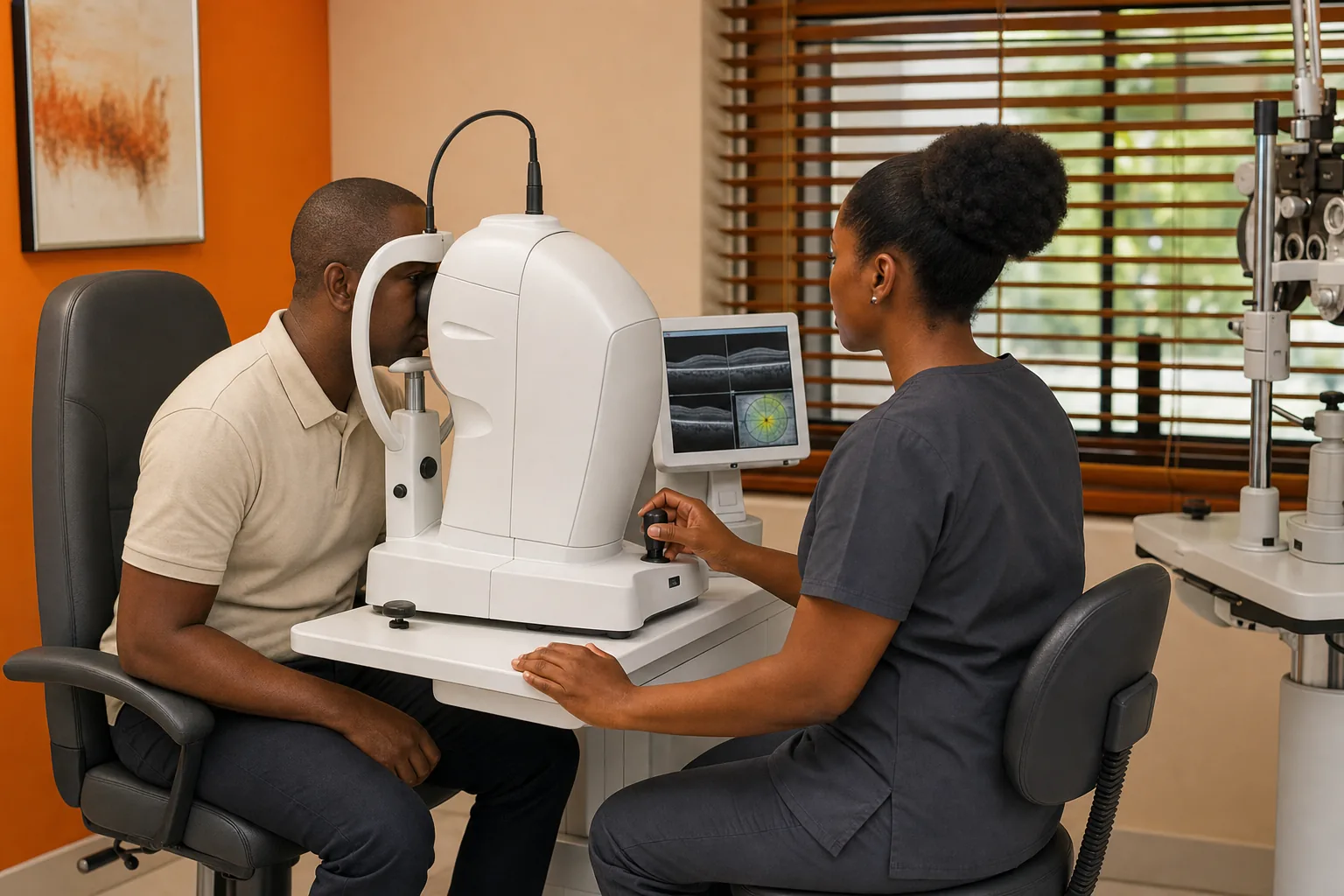
Testing can identify risk before you notice a missing patch of side vision. It cannot restore sight already lost, so early review matters. Our goal is to protect the vision you still have through education, monitoring, and appropriate care.
Key Takeaway:A glaucoma assessment needs several tests because pressure alone cannot confirm or exclude the disease.
When to Arrange Glaucoma Testing Near You
If you’re searching for glaucoma testing near me because of a new symptom, don’t wait for a routine visit. Sudden eye pain with blurred vision, headache, nausea, or vomiting can signal angle-closure glaucoma. Seek urgent medical care.
Most glaucoma develops slowly. The open-angle form may cause no clear symptom until side vision has already changed. This makes routine testing important for people who feel perfectly well.
Arrange an assessment if you have:
A parent, sibling, or child with glaucoma.
Diabetes or high blood pressure.
A history of eye injury or inflammation.
Very strong short-sightedness or long-sightedness.
Previous eye surgery linked to a change in pressure.
New trouble with side vision or frequent changes in glasses.
Age also affects risk. Many people first need closer glaucoma checks in adult life, but there is no single schedule for everyone. Your ophthalmologist may set the interval based on your pressure, optic nerve appearance, family history, and past results.
A routine eye exam may find a concern. That does not always mean you have glaucoma. It may mean you need more detailed tests or a repeat measurement under different conditions.
For example, a pressure reading can vary during the day. Corneal thickness can also affect how a pressure result should be understood. One reading gives useful information, but it does not tell the whole story.
Diabetes deserves extra care. It can affect the retina and may occur alongside glaucoma. EyesTT also provides diabetic eye care in Trinidad and Tobago , which can help keep related eye checks within one care plan.
We understand that arranging an appointment can be hard when you work, care for family, or travel between communities. Before booking, ask how long the visit may take and whether more than one test can happen during the same appointment.
Wait times and accepted insurance plans aren’t listed consistently in the available local information. Ask the clinic directly about both points. You should also ask whether your referral, medical history, or old test reports are needed.
Bring a list of medicines and eye drops. Include drops prescribed for another condition. Some medicines can affect eye pressure or interact with glaucoma treatment.
Bring your glasses too. If you have had previous scans or visual field tests, those records help us compare change over time.
Regular testing is especially useful after a result described as ocular hypertension. That term means pressure is higher than expected without clear optic nerve damage. Some people remain stable, while others later show changes. Monitoring helps separate those paths.
The right time to book is before vision loss becomes part of daily life. If symptoms appear suddenly, treat that as urgent rather than waiting for a standard appointment.
What Happens During a Glaucoma Eye Examination
A glaucoma eye examination usually starts with questions, then moves through a set of painless tests. The order can change based on your symptoms, age, medical history, and first findings.
We begin by asking about your sight. We may ask if you bump into objects, miss things at the side, see halos, or notice a change in one eye.
Next, we check visual acuity. You read letters or symbols at set distances. This does not diagnose glaucoma by itself, but it shows how clearly you see at the time of testing.
Tonometry measures IOP. A device may gently touch the front of the eye after numbing drops. Another method can estimate pressure without contact. The test takes little time.
Try to keep your forehead and chin still. Stay relaxed. Squeezing your eyelids or holding your breath can make the test harder to perform.
A dilated examination may follow. Drops widen the pupil so we can inspect the retina and optic nerve. You may notice glare or blurred near vision for part of the day.
Funduscopy uses magnification and light to inspect the back of the eye. We look at the optic disc, blood vessels, retina, and retinal nerve fibre layer. The optic disc is the point where the optic nerve leaves the eye.
OCT adds a layer-by-layer image. You rest your chin on the machine and look at a target. Nothing touches the eye. The scan can show patterns in the nerve tissue that need review or follow-up.
Visual field testing checks how much you see while looking straight ahead. One eye is tested at a time. You press a button when you see lights, even when the light seems faint.
This test can feel tiring. Keep looking at the central target. Don’t press for every guess. The result is more useful when it reflects what you truly saw.
Gonioscopy examines the drainage angle. Numbing drops are used first. A special lens rests on the eye for a short time while the ophthalmologist checks whether the angle is open, narrow, or closed.
Pachymetry may also be used to measure corneal thickness. This helps the doctor judge how much weight to place on an IOP reading.
Test | What it checks | Why it may matter |
Tonometry | Pressure inside the eye | High pressure can raise risk, but it does not confirm glaucoma alone. |
OCT | Retinal and optic nerve structure | Shows tissue patterns that can be compared over time. |
Visual field test | Central and side vision | May show functional loss linked to optic nerve damage. |
Gonioscopy | Drainage angle | Helps identify open-angle or angle-closure features. |
Funduscopy | Retina and optic nerve appearance | Lets the doctor inspect the back of the eye directly. |
Pachymetry | Corneal thickness | Adds context to pressure readings. |
The clinical overview of glaucoma tests explains that doctors use these tests together. Each one answers a different question. A result that looks unusual may lead to another test rather than an immediate diagnosis.
Testing is usually safe. Dilation may leave you sensitive to light, so sunglasses can help. If your vision stays blurred, arrange someone else to drive.
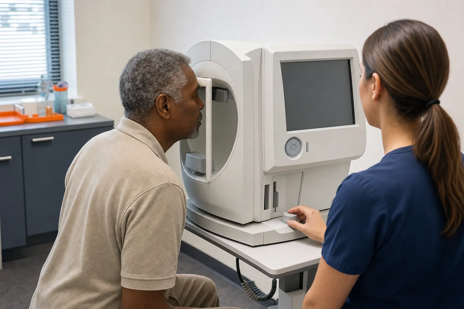
Most people receive an explanation during the appointment. Ask us to repeat any term that feels unclear. You should leave knowing what was tested, what the findings suggest, and what happens next.
How Doctors Interpret Eye Pressure and Other Results
When reviewing glaucoma testing near me results, doctors look for a pattern across both eyes. A single pressure number rarely gives enough information on its own.
IOP is one part of the assessment. Higher pressure can increase the chance of optic nerve damage. Yet some people with high pressure never develop glaucoma, while others develop damage at pressure that appears ordinary.
Doctors also study the optic nerve. They look at its shape, colour, rim tissue, and the cup at its centre. A large cup may be normal for one person. A change over time can be more concerning.
OCT shows the retinal nerve fibre layer and nearby structures. If the scan shows thinning, its findings can be considered alongside the appearance of the nerve and the visual field. The scan needs clinical interpretation because normal anatomy varies between people.
The visual field shows how the eye functions. A person may have a structural change without a matching field loss. That can lead to closer checks. A repeat test may be needed if the first result was unreliable.
Common reasons for an unreliable visual field include tiredness, poor focus, incorrect instructions, or pressing the button too often. One weak test should not define your care plan.
Gonioscopy helps identify the type of glaucoma risk. A narrow or closed angle can change the urgency and treatment discussion. It can also show whether the drainage area is open enough for certain treatments.
Corneal thickness adds context to pressure. A thicker or thinner cornea can affect how a pressure reading is understood. It does not replace the other tests.
Doctors may describe you as:
Glaucoma suspect:One or more findings raise concern, but the diagnosis is not confirmed.
Ocular hypertension:Pressure is raised without clear evidence of optic nerve damage.
Glaucoma:The findings support optic nerve damage, with or without raised pressure.
Stable glaucoma:Current results do not show meaningful change from earlier records.
Progressing glaucoma:Tests suggest that damage or field loss is changing over time.
These terms describe a working clinical picture. They are not a prediction of exactly how your sight will change.
Future results can be compared with your first set of measurements. That is why keeping records matters. A pressure that stays similar may still need attention if the field or OCT changes.
Early glaucoma may cause no symptoms, so testing can help identify changes before they are noticed.
Ask these questions when results are discussed:
Is there evidence of optic nerve damage?
Is the visual field reliable?
Does one eye differ from the other?
Do we need a repeat test?
When should the next review happen?
What warning symptoms need urgent care?
Good care includes clear answers. You should understand the finding before deciding on the next step.
How to Choose a Local Glaucoma Testing Clinic
Choosing a clinic for glaucoma testing near me starts with scope. Ask whether the service can assess pressure, optic nerve structure, visual field, drainage angle, and the retina.
A clinic that can arrange several tests may reduce delays between separate appointments. It may also make it easier to compare results because the records stay within one care pathway.
Ask what happens if a test raises concern. Will an ophthalmologist review the findings? Can the clinic arrange follow-up? Is treatment planning available if glaucoma is confirmed?
EyesTT provides ophthalmology and eye care in Trinidad and Tobago. Its glaucoma diagnostic service lists OCT, visual field perimetry, IOP measurement, gonioscopy, and funduscopy. EyesTT also provides broader eye exams, diagnostic services, surgery, and optical products.
We keep the discussion focused on your needs. A person with a family history may need a different review plan from someone with sudden symptoms. A child also needs age-appropriate assessment.
Ask about preparation before you arrive. Find out if you should bring old records, use your usual drops, or allow time for pupil dilation.
Ask about usable matters too:
How long should you allow for the visit?
Could dilation affect your ability to drive?
When will the doctor discuss your results?
What are the fees for each test?
Which insurance plans or payment options are accepted?
What happens if more testing is needed?
Available local sources do not provide standard wait times or accepted insurance details. That gap matters. Clear answers help patients plan work, transport, childcare, and follow-up.
Check that the clinic explains what each test means. Equipment alone doesn’t make a diagnosis. The value comes from skilled review and a plan that matches the findings.
For some patients, treatment may later include drops, surgery, or laser therapy. EyesTT’s Tango Neo SLT/YAG Laser page explains its glaucoma and cataract laser treatment scope. EyesTT does not provide laser refractive surgery, such as LASIK. Its listed laser work is focused on SLT/YAG and Easyret retinal laser services.
That distinction can help you ask a better question: does the clinic provide the care your eye condition needs? Glaucoma care does not require a clinic to offer every kind of eye procedure.
Choose a service that makes the next step clear. If the clinic cannot explain what happens after an abnormal result, keep asking before you book.
What Happens After Testing: Monitoring and Treatment Planning
After glaucoma testing, the ophthalmologist reviews the full set of findings. You may receive reassurance, a request for repeat testing, a monitoring plan, or a treatment recommendation.
If the findings are normal, you may still need future checks based on your risk. A normal result today does not remove family history, diabetes, age, or past injury from your health record.
If you are a glaucoma suspect, the next step may be closer monitoring. The doctor may repeat IOP, OCT, or visual field testing. The aim is to see whether a finding stays stable or changes.
If glaucoma is confirmed, treatment usually aims to lower pressure and protect the optic nerve. Treatment may include prescription eye drops, laser therapy, or surgery. The right choice depends on the type of glaucoma and the amount of damage.
Eye drops work in different ways. Some reduce fluid production. Others help fluid leave the eye. Use them exactly as prescribed, even when your eyes feel fine.
Tell us if drops cause redness, stinging, blurred vision, breathing problems, or other concerns. Don’t stop a prescribed treatment without speaking with your eye-care team.
SLT, or selective laser trabeculoplasty, can help fluid drain in some open-angle glaucoma cases. YAG laser treatment has different uses, including certain problems after cataract surgery. Your ophthalmologist will explain which procedure fits your diagnosis.
Surgery may be discussed when pressure stays too high despite other care. The conversation should cover the aim, expected follow-up, possible risks, and what you must do after the procedure.
Monitoring is part of treatment. At each visit, we may compare pressure with earlier readings. We may also review OCT images, visual fields, and the optic nerve.
Keep every appointment, even when your sight seems unchanged. Glaucoma can progress quietly. Regular review gives the care team a chance to respond before a small change becomes a larger loss.
Bring your eye drops to follow-up visits. Tell us how often you use them. If cost, forgetfulness, hand strength, or side effects make the plan hard, say so. We can only adjust the plan when we know what is getting in the way.
Family members may need testing too. Share the diagnosis with close relatives and encourage them to arrange an eye exam. A family history can affect their risk.
Seek urgent care for sudden severe eye pain, a sudden drop in vision, halos with nausea, or a red painful eye. Those symptoms need prompt assessment rather than a routine follow-up.
Pro Tip:Keep a simple record of your test dates, pressure readings, medicines, and follow-up plans. It helps you spot missed visits and gives your care team useful context.
At EyesTT, our goal is complete eye care that supports your quality of life. We explain the findings in plain language and help you understand the next decision. That is part of taking eye care into the future.
FAQ
What does glaucoma testing include?
Glaucoma testing can include IOP measurement, OCT, visual field testing, gonioscopy, and funduscopy. An ophthalmologist may also check corneal thickness and dilate your pupils. The tests look at pressure, optic nerve structure, side vision, fluid drainage, and the retina. EyesTT lists these five core glaucoma diagnostic methods.
Can I have glaucoma with normal eye pressure?
Yes, glaucoma can occur even when eye pressure appears normal. Doctors also look for optic nerve damage and visual field changes. A normal IOP reading does not rule out the disease. That is why glaucoma testing near me should involve more than a pressure check when risk or symptoms are present.
Is glaucoma testing painful?
Most glaucoma tests are quick and painless. Numbing drops may be used for some pressure or angle tests. Dilation can cause temporary glare and blurred near vision. Visual field testing may feel tiring because you must keep looking at a central target. Ask about transport if your pupils will be dilated.
When should I get glaucoma testing?
You should arrange testing if you have a family history, diabetes, high eye pressure, past eye injury, or a change in side vision. Sudden eye pain with blurred vision, headache, nausea, or vomiting needs urgent medical care. Your ophthalmologist can set a review schedule based on your results and risk.
How long does a glaucoma eye exam take?
The time varies because glaucoma testing may involve several exams. Dilation, OCT, visual field testing, and a doctor review can make the visit longer than a basic sight test. Ask EyesTT when booking how much time to allow. Also ask whether you should arrange transport after dilation.
What happens if my glaucoma test is abnormal?
An abnormal result does not always confirm glaucoma. Your ophthalmologist may repeat a test, compare both eyes, or review earlier scans. If the pattern supports glaucoma, the doctor may recommend monitoring, eye drops, laser treatment, or surgery. You should receive a clear follow-up plan before leaving.
Conclusion
Choose glaucoma testing that looks at the whole eye, not pressure alone. EyesTT provides five core glaucoma diagnostic methods alongside broader eye care in Trinidad and Tobago. If you have a risk factor or new concern, arrange an ophthalmology appointment and bring your medicines and past eye records. We’re here to help you understand each result and plan the next step.


Comments